
Our research concerns dosimetry by electron paramagnetic resonance (EPR), thermoluminescence (TL) , UV-Vis spectrophotometry and Nuclear Magnetic Resonance (NMR) and image analysis acquired by means of advanced Magnetic Resonance Imaging (MRI).
The dosimetry is aimed at quantifying the absorbed dose due to ionizing radiation through measurements of the variations of physical, chemical and biological properties of the materials after irradiation.
 The main research is aimed at the study of new materials and additives used for the realization of ESR solid state dosimeters alternative to those widely distributed of alanine (e.g. ammonium tartrate and gadolinium has been studied in order to increase the sensitivity to both gamma photons and thermal neutrons). This research is supported and aided by the development and the implementation of Monte Carlo codes able to predict the response of ESR solid state dosimeters after irradiation by different radiation beams and with different geometries. In particular, analysis techniques have been developed and allow to obtain information on the ionizing linear energy transfer (LET) and indirectly on the relaxation properties (relaxation times T1 and T2) in samples exposed to radiation of different LETs. Furthermore, research activities in retrospective dosimetry are carried out in order to develop techniques able to provide rapidly doses in case of radiological emergency. | 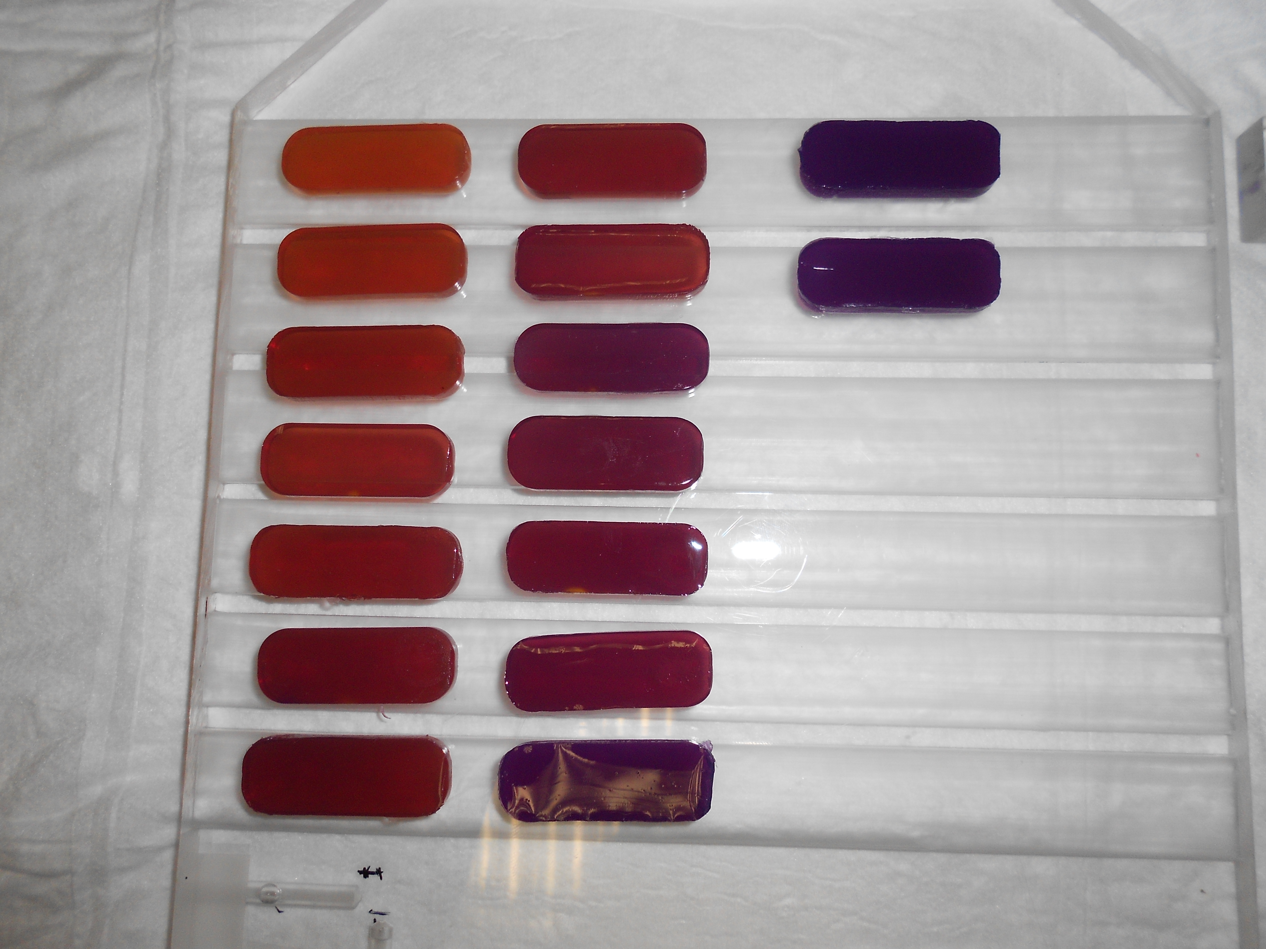 Use of ionizing radiation in the clinical filed has stimulated and increased development and use of numerous dosimetric devices aimed at providing values of the absorbed dose as a result of therapy or radiodiagnostic examinations as accurate as possible. There is a special interest in dosimetric gels since they can have as an advantage the ability to provide even a 3D dose distribution if suitably prepared as phantoms. The dosimetric gels are constituted by a gelatinous tissue-equivalent matrix, in which are dissolved chemical radiation-sensitive species that after irradiation are modified as a function of absorbed dose. In our research measurements using NMR relaxometry and UV-Vis spectrophotometry of gel Fricke dosimeters (FXG) are performed. | 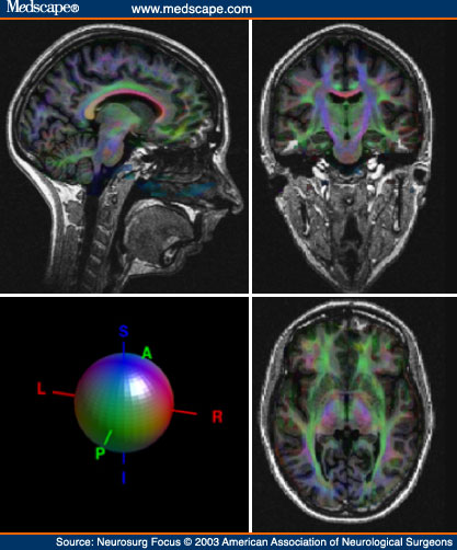 In medical imaging the nuclear magnetic resonance imaging (MRI) assumes a great importance because this technique has a better contrast than other structural tomographic techniques, such as computed tomography (CT), and does not make use of ionizing radiations. One of the key features of the MRI images is their quantitative aspect. Our reaserch is aimed at developing techniques and software to analyze images obtained by means of the advanced MRI techniques listed below:
|