Confocal
and Two Photon Microscopy Laboratory
University
laboratory
Scientific
responsible: Prof. V. Militello
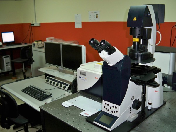
Confocal microscopy is an optical imaging technique, widely diffused as a tool for a wide number of studies in Biophysical and Biomedical research as well as in material sciences. It allows to obtain high resolution optical images both of fixed and living fluorescent specimens. This technique presents several advantages with respect to conventional microscopy due to an increased micrograph contrast and to the reduction of out-of-focus light or flare contributions in specimens that are thicker than the focal plane. This result is achieved by means of a confocal "pinhole" situated in front of the image plane which acts as a spatial filter and allows only the in-focus portion of the light to be imaged. Confocal microscopy also allow to non invasively collect optical sections (0.5-1.5 micron) from thick specimens, this feature together with recent advances in fluorescent proteins and dyes technology have allowed to monitor biological processes in live cells in 3D and in real time.
Two-photon excitation microscopy is a fluorescence imaging technique as well, like confocal microscopy it provides thin optical sections from deep within thick, scattering specimens. For this technique fluorophore excitation (and thus emission) occurs only at the focal plane of the microscope this feature spatially confine two-photon excitation giving rise to several advantages especially in 3D imaging. Moreover penetration depth of the excitation beam is increased and since the pinhole is not necessary in the detection path fluorescence collection efficiency is increased. Moreover, the localisation of excitation volume of two-photon excitation markedly reduces overall photobleaching and photodamage.
Both confocal and two photon excitation microscopy allow to measure FRET (Foster Resonance Energy Transfer) in the images. FRET is a useful physical phenomenon to measure molecule distance, and has emerged as a method to detect intermolecular interactions or protein conformational changes in living cells. FRAP (Fluorescence Recovery After Photobleaching ) can be also measured, FRAP measurements provide information about the mobility of a fluorescent molecule in a defined compartment. This thecnique can be used to obtain suitable information on diffusion of proteins or proteins complexes in model matrices or in living cells
The high spatial resolution, low photobleaching and spectral discrimination inherent of these two fluorescence imaging techniques result appealing also for the study of luminescent point defects in silica.
Policy
for external users (in italian)
|
Confocal
Microscopy Setup
|