Raman and Micro-Raman Laboratory
CNR-IBF Laboratory - Technological
Transfer Project "Oil
Olive Quality Control" - POR
Sicilia - Mis. 3.15-C
Laboratory directed by: Dr.
Simone Agnello
Raman spectroscopy is used to study the vibrational modes of biological samples directly in aqueous systems, since the vibrations of H2O are not Raman-activated. In addition, Raman is usually used to characterize and to study the variations in the protein polarity in the surrounding of Tyrosine residues, by a Raman doublet in the 820860 cm-1 interval. Moreover, it is possible to monitor the S-S conformation through the analysis of a broad band at about 510 cm-1, giving information on the different configurations of the CβSSCβ disulfides bridges (ggg, ggt, ) and by the spectral changes visible in the region due to νCS modes of Cys. Two different Raman spectrometers are available in the lab. Their main technical features are reported below.
|
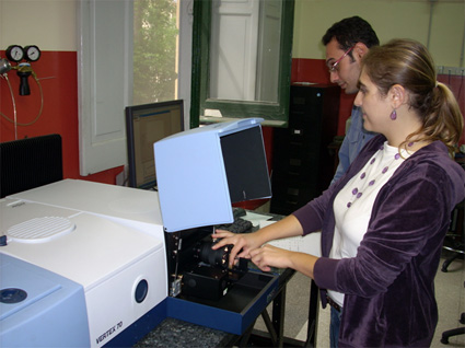
FT-Raman: Bruker VERTEX-70 RAMII
|
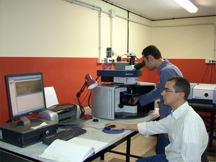 |
Micro-Raman: Bruker SENTERRA |
This laboratory is integrated in MeSIAM - Regional Laboratory of CNISM |
||